Few sights are as alarming to a French Bulldog parent as waking up to find a bright red, swollen, cherry-sized mass protruding from the inner corner of their dog’s eye. Known clinically as a prolapsed gland of the third eyelid, or simply “cherry eye,” this condition is an unfortunately common genetic challenge within the Frenchie breed.
When a cherry eye flares up, the immediate reaction of many first-time owners is panic. The swelling looks painful, angry, and unsightly. You are suddenly faced with a massive veterinary decision: Do you have the gland surgically cut out (excision)? Or do you have it tucked and sewn back into place (the Morgan Pocket Suture method)?
Related Reading: Training & Behavior | Grooming & Care | French Bulldog Colors
In my ten years of breeding and showing champion French Bulldogs, I have seen this genetic trait manifest across various bloodlines. I have guided numerous puppy families through the confusing veterinary options, weighed the long-term biological consequences of dry eye, and tracked surgery recurrence rates across multiple clinical methods.
This guide is designed to deconstruct the anatomy of cherry eye, compare the pros and cons of the two primary surgical interventions, analyze their true recurrence rates, and give you a practical, breeder-tested roadmap for postoperative recovery.
1. The Anatomy of Cherry Eye: Why Are Frenchies Genetically Vulnerable?
To make an informed surgical decision, you must first understand the anatomy of a Frenchie’s eye. Unlike humans, dogs possess a third eyelid (nictitating membrane) located in the inner corner of the eye, beneath the lower eyelid. This third eyelid serves as a critical protective shield, sweeping across the cornea like a windshield wiper to clear away dust, debris, and allergens.

Tucked safely inside the base of this nictitating membrane is the nictitans gland. This gland is a powerhouse; it is responsible for producing approximately 30% to 50% of the entire aqueous tear film that keeps your dog’s eye lubricated, nourished, and free from corneal ulcers.
In healthy dogs, the gland is anchored firmly to the orbital rim of the eye by a thick band of fibrous connective tissue. However, because French Bulldogs are bred with a compact, shortened skeletal structure (brachycephaly), they frequently inherit weak, loose, or malformed connective tissue anchors.
When this fibrous anchor is too weak to hold the gland down, the gland slips out of its pocket and pops up over the margin of the third eyelid. Once exposed to the dry outside air, the gland quickly becomes inflamed, congested, and swollen, transforming into the classic red “cherry” mass.
2. Excision (Surgical Removal): The Outdated Quick Fix
In the past, the standard treatment for cherry eye was incredibly simple: the veterinarian would apply local anesthesia and cut the prolapsed gland out entirely. This procedure, known as gland excision, is fast, highly cost-effective, and guarantees a 0% recurrence rate—after all, you cannot prolapse a gland that no longer exists.
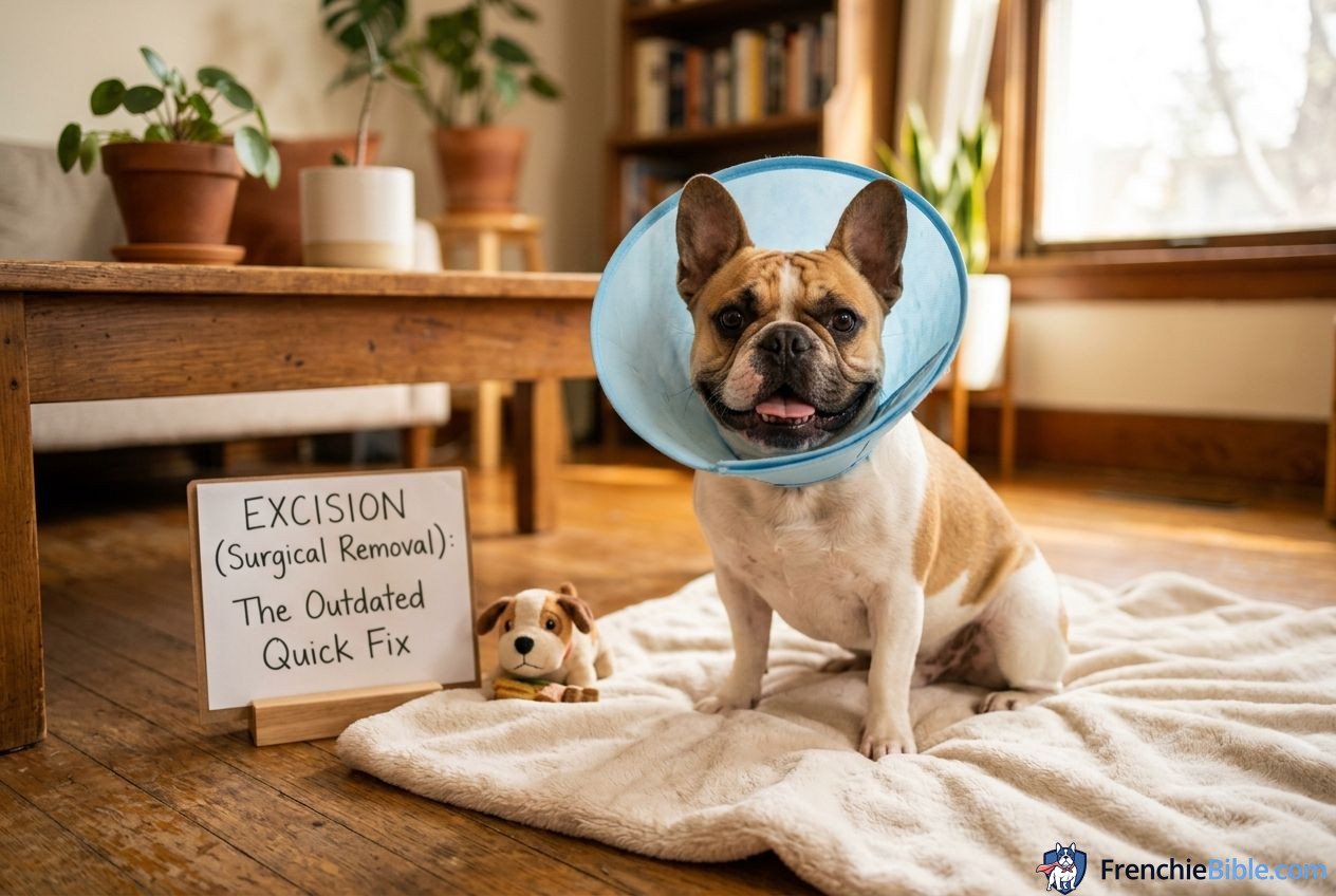
The Hidden Danger of Excision: Dry Eye (KCS)
As a preservationist breeder, I must issue a stern warning: Excision is an outdated, dangerous quick fix that should be avoided at all costs, except in rare oncology cases.
When you cut out the nictitans gland, you are permanently destroying up to half of your Frenchie’s tear-producing capacity. While a young Frenchie’s eye may seem fine immediately after excision, their remaining tear glands will slowly struggle to keep up as they age.
Within 2 to 5 years post-excision, a massive percentage of these dogs develop Keratoconjunctivitis Sicca (KCS), commonly known as chronic dry eye.

Without natural lubrication, the cornea becomes chronically dry, irritated, and prone to painful corneal ulcers. Over time, to protect itself, the cornea will deposit dark pigments over the surface (pigmentary keratitis), slowly blinding your dog.
If your Frenchie undergoes excision, you will likely spend the next 10 to 12 years of their life applying expensive immunosuppressant eye drops (like Cyclosporine or Tacrolimus) and artificial tears 3 to 4 times a day, costing you thousands of dollars and causing your dog chronic discomfort.
3. The Pocket Suture Method (Morgan Pocket): The Preservation Standard
Modern veterinary ophthalmology has shifted completely toward gland preservation. Instead of removing the gland, surgeons use specialized techniques to tuck the gland back into its natural anatomical position and secure it.
The most widely accepted and successful preservation technique is the Morgan Pocket Suture method.
How the Morgan Pocket Method Works
During this surgery, the veterinarian makes two small, shallow incisions in the conjunctiva (the pink inner lining of the third eyelid) directly above and below the prolapsed gland.
The surgeon gently pushes the inflamed gland down into this newly created tissue pocket. Using an incredibly fine, dissolvable suture material, the surgeon sews the two tissue flaps together over the gland, sealing it safely inside the pocket.

The ends of the suture are tied on the outside surface of the third eyelid, facing away from the eye, to prevent the knots from rubbing against and scratching your Frenchie’s sensitive cornea.
The Benefits
- Preserves Natural Tear Production: By saving the gland, your Frenchie retains 100% of their natural tear film capacity, preventing KCS and safeguarding their vision for life.
- Maintains Eye Structure: The third eyelid retains its natural shape and sweeping motion, keeping the eye clear of daily irritants.
4. Comparing the Methods: Recurrence Rates, Costs, and Recovery
While gland preservation is the gold standard, it is not without its challenges. The primary hurdle is the recurrence rate—the probability that the surgical tuck will fail and the gland will pop out again.
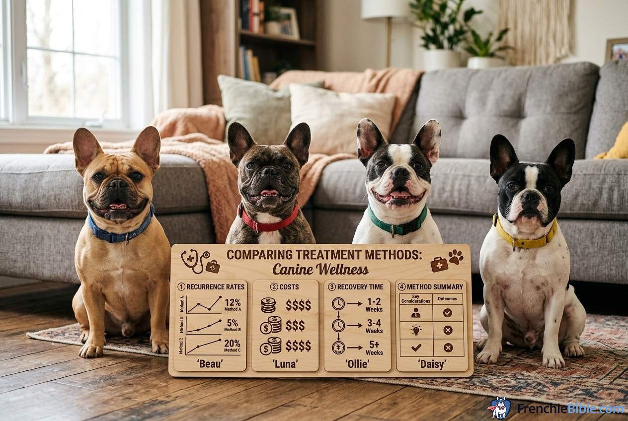
Recurrence Rates Analyzed
Because French Bulldogs have massive facial muscle tension and breed-specific orbital pressure, the pressure pushing against the surgical pocket is intense.
- Excision Method: 0% Recurrence. (But leads to dry eye).
- Pocket Suture Method: 10% to 20% Recurrence.
In my breeding history, I have found that if a pocket suture is going to fail, it almost always fails within the first 14 to 30 days post-surgery, during the active healing phase when the dissolvable sutures are weakening but the tissue has not yet formed strong scar tissue. If the pocket holds past day 45, it is highly likely to hold for the rest of your Frenchie’s life.
The Primary Factors That Drive Surgical Failure:
- Gland Inflammation Size at Time of Surgery: If the gland is allowed to remain prolapsed for months before surgery, it becomes chronically swollen, enlarged, and fibrotic. Trying to stuff an oversized, angry gland into a small conjunctival pocket is like trying to close an overstuffed suitcase—the sutures will likely rip through the tissue.
- Lack of Postoperative Compliance: If the dog is allowed to rub their face against carpets or scratch their eye with their paws, they will rip the delicate sutures open instantly.
- Surgeon Experience: Placing sutures in a membrane thinner than a paper towel requires immense precision. General practitioners have higher failure rates than board-certified veterinary ophthalmologists.
5. Postoperative Recovery SOP: A Breeder’s Guide to Zero Complications
As a breeder, I have learned that the success of a cherry eye tuck is 50% surgeon skill and 50% home recovery care. If you do not control your Frenchie’s behavior during the critical 14-day healing window, the surgery will fail.
Here is my non-negotiable postoperative care protocol:
Step 1: The Hard E-Collar (No Exceptions)
Do not use soft, inflatable “donut” collars. Frenchies are short, stocky, and highly flexible; they can easily bend their necks around an inflatable collar and rub their eye against the corner of a coffee table or a couch cushion.
- Rule: Your Frenchie must wear a rigid, plastic Elizabethan collar (cone of shame) for a full 10 to 14 days post-surgery. The cone should only be removed under direct, hand-held supervision for cleaning, and put immediately back on.

Step 2: Strict Activity Restriction (Crate Rest)
Every time your Frenchie barks, runs, jumps, or pants heavily, the blood pressure inside their head and eyes spikes. This increased orbital pressure pushes directly against the delicate suture line.
- Rule: Keep your Frenchie in a comfortable, quiet crate or a small playpen. No running, no jumping onto sofas, and absolutely no rough play with other household pets. Leash walk them strictly for short bathroom breaks only.
Step 3: Zero Neck Pressure (Harness Only)
Never attach a leash to a neck collar after cherry eye surgery. A collar pulls directly on the jugular veins, increasing intracranial and intraocular pressure, which can tear the sutures.
- Rule: Use a wide, padded Y-harness that distributes weight entirely across the chest and shoulders, completely bypassing the neck.
Step 4: Master the Eye Drop Technique
You will be sent home with antibiotic drops (such as Tobramycin) and anti-inflammatory drops (such as Flurbiprofen or Prednisolone acetate).
- Rule: Always wash your hands thoroughly before applying drops. Approach your Frenchie from behind or above their head rather than coming directly at their face, which triggers a panic-blink reflex. Gently pull the upper eyelid upward with your thumb and let the drop fall onto the sclera (white part of the eye) without letting the plastic dropper tip touch the eyeball.
6. Frequently Asked Questions (FAQ)
Q1: Can I just leave my Frenchie’s cherry eye untreated if it doesn’t seem to bother them?
No, leaving a prolapsed gland untreated is highly discouraged. While it may not seem painful in the early stages, the exposed gland is no longer lubricated by tears. Over time, it will swell, dry out, scab, and become a breeding ground for bacterial infections. Additionally, a chronically prolapsed gland will lose its ability to produce tears due to glandular tissue death, leading to KCS (dry eye) even without surgical excision.
Q2: If cherry eye occurs in one eye, will it inevitably happen in the other?
In the French Bulldog breed, cherry eye is highly bilateral. There is an approximate 60% to 70% chance that if one eye prolapses, the other eye will prolapse within the next 3 to 12 months. This is because the underlying genetic connective tissue weakness exists in both eyes. If your Frenchie requires surgery for one eye, discuss with your veterinary ophthalmologist whether a preventative “tuck” or a combined procedure is appropriate if the second eye is already showing signs of looseness.
Q3: How long should I wait after the cherry eye pops up before scheduling the surgery?
You should aim to have the surgery performed within 2 to 4 weeks of the initial prolapse. You want to give the eye a few days to calm down with the help of anti-inflammatory drops prescribed by your vet, but you must not wait so long that the gland becomes permanently enlarged, fibrotic, or damaged by air exposure. A healthy, fresh prolapse has a significantly higher surgical success rate than a dry, hard mass that has been exposed for six months.
Q4: My Frenchie is rubbing their cone against the carpet, and the eye looks slightly bloody. What should I do?
A small amount of pink-tinged tear discharge is normal for the first 2 to 3 days after surgery. However, if your Frenchie is actively rubbing the cone against surfaces, they are transferring kinetic energy directly to the eye, which can tear the stitches. You must immediately restrict their movement further (using a smaller crate) or use a “double-coning” method (placing a soft donut collar underneath the hard plastic cone to completely limit neck movement and keep the cone off the floor). If you see bright red blood or notice a sudden gap in the eye corner, contact your surgeon immediately.
Q5: Can I prevent cherry eye through diet, vitamins, or eye massage?
Unfortunately, because cherry eye is a structural, genetic defect in the strength of the connective tissue anchor, it cannot be prevented by dietary changes, joint supplements, or vitamins. Some owners attempt to “massage” the gland back into place when it first pops up. While this can temporarily slide the gland back down, it is not a permanent cure; the weak anchor will eventually allow it to slip out again during the next bout of excitement, sneezing, or play.
7. Disclaimer
This guide is based on my ten years of hands-on experience raising, breeding, and caring for French Bulldogs. I am not a veterinarian, and this content is purely for educational and supportive purposes. Cherry eye is an ocular condition that requires professional clinical assessment. If your Frenchie is showing signs of eye pain, squinting, heavy discharge, or a clouding cornea, please consult a licensed veterinarian or a board-certified veterinary ophthalmologist immediately for a proper diagnosis and treatment plan.